Percutaneous Nephrostomy
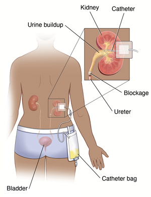
Percutaneous nephrostomy is a procedure where a small tube (catheter) is put through your skin into your kidney to drain your urine. This procedure is done by a specially trained healthcare provider called an interventional radiologist.

Why percutaneous nephrostomy is done
Percutaneous nephrostomy may be needed when a kidney or a tube (ureter) leading from a kidney to your bladder gets blocked. This can happen because of kidney stones, tumors, or another cause. The blockage can cause a backup of urine in the kidney. This procedure is done to stop pain, infection, and kidney damage.
Risks of percutaneous nephrostomy
All procedures have some risks. Possible risks of this procedure include:
-
Bleeding into or around your kidney or damage to your kidney
-
Blockage of the catheter
-
Blood infection (sepsis)
-
Kidney infection
-
Problems because of the X-ray dye (contrast medium). These include allergic reaction or kidney damage.
-
Skin infection around the catheter site
-
The catheter may need to be replaced if it is used for a long time. The same procedure is used.
-
Urine leak
-
X-ray radiation exposure, which is considered to be low level and safe
Getting ready for your procedure
Tell your healthcare provider if you:
Tell your healthcare provider about any recent illnesses, all medical conditions, and all medicines you take. You may need to stop taking some or all of them before your procedure. This includes:
-
All prescription medicines
-
Any street drugs you may use
-
Herbs, vitamins, and other supplements
-
Over-the-counter medicines that don’t need a prescription, including aspirin and ibuprofen
Also be sure to:
-
Follow any directions you’re given for not eating or drinking before your procedure.
-
Follow any other directions from your healthcare provider.
-
Plan to have a relative or friend drive you home after your procedure. You can’t drive yourself.
During your procedure
-
You will change into a hospital gown.
-
An IV (intravenous) line is put into your hand or arm to give you fluids and medicines. You will then lie on your stomach on an X-ray table. You may be given medicine to help you relax and make you feel sleepy.
-
The skin on your lower back is numbed with an injection of local anesthesia.
-
The radiologist will use CT scan, ultrasound, or fluoroscopy, images as a guide. They will insert a needle through your lower back into your kidney. X-ray dye may be injected through this needle into your kidney. This fluid makes your kidney easier to see on X-ray images. The X-ray images can show exactly where your kidney or ureter is blocked.
-
The needle is then replaced with a thin tube called a drainage catheter. The catheter is attached to a drainage bag. This bag collects the urine that drains from your kidney. The catheter may be stitched (sutured) or taped to your skin. This helps keep it in place and stop it from moving.
-
The entire procedure takes about 1 to 2 hours. You will remain in the recovery room until you are completely awake and ready to go home.
After your procedure
The catheter will stay in place until the problem that caused the urine buildup is treated. This may be for as little as a day or as long as a few weeks or months. The bag is secured to your leg so you can walk around. During the time the catheter is in place you should:
-
Keep the skin around the catheter clean and dry.
-
Be careful not to move or knock the catheter out of place. Make sure that the drainage bag is secured firmly to your leg.
-
Empty the drainage bag often. This keeps the weight of the bag from pulling on the catheter.
-
Your healthcare provider will give you detailed directions on how to care for your catheter and drainage bag.
When to call your healthcare provider
Call your healthcare provider if your urine becomes cloudy or smells bad, or if you develop fever or chills. Follow all directions given to you by your healthcare provider.